
Introduction to cherry eye

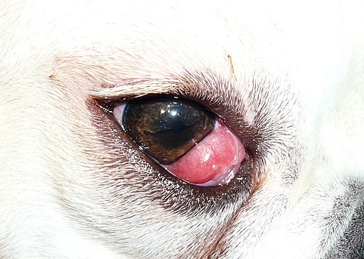
A cherry eye is a non-life-threatening condition that occurs in dogs, and less often in some cat breeds. It is an extremely descriptive term, as one can see an oval, bright red swelling in the inside corner of an affected dog’s or cat’s eye, resembling a cherry. As a pet owner one can easily become quite alarmed by seeing this, but fortunately, it only causes slight irritation to the dog initially and you will have time to attend to it and take your animal to the vet before the condition gets out of hand. It is never a good idea to just leave it be. The condition tends to occur more commonly in younger dogs and cats, usually between the ages of 2 and 6 years.
How does it happen that an animal develops a cherry eye?
A cherry eye is in actual fact a protrusion (or bulging out) of the gland of what is colloquially called the third eyelid. Dogs and cats have three eyelids, the top and bottom lids that close up and down over the eyeball as in humans, and then a third eyelid, otherwise called the nictitating membrane underneath the upper and lower eyelids. If you press on the eyeball through the upper eyelid, you will notice the third eyelid moving across the ball of the eye from the inside corner of the eye towards the outside corner of the eye. This eyelid contains a gland that produces up to 30% of the tear production of the eye. The third eyelid provides extra protection to the animal’s eye and keeps the eye moist. The gland in the nictitating membrane is anchored to the corner of the eye by a connective tissue band. For reasons unknown, this connective tissue starts to weaken and the gland slips out of its pocket. If this happens, the gland is exposed to sun, wind, dust, and trauma from the outside. The gland becomes red and swollen, and eventually painful, due to inflammation. One or both eyes may be affected at the same time. The most common breeds affected by this condition are Beagles, Bulldogs, Spaniels, Shih Tzus, Pekingese, and other brachiocephalic (flat-faced) breeds. The condition is rare in cats but Burmese and Persians seem to have a higher incidence of cherry eye.
Other clinical signs associated with cherry eye.
You will quickly notice the red swelling in the corner of your animal’s eye. Other signs that you might notice are a mucoid discharge from the eye and/or redness in the tissue surrounding the eye, called conjunctivitis. Your pet might also show you he/she is experiencing discomfort by pawing at the eye or rubbing his/her face against objects. This can cause even more trauma to the exposed gland.
Course of action with cherry eye
Due to the fact that some dogs don’t seem phased by the popped out gland, some owners might opt to leave it like that. Not treating the gland may however cause more serious problems to the affected eye in years to come. As more damage is inflicted onto the popped out gland, the amount and quality of tear film that protects the eye will decrease causing chronic inflammation and irritation to the eye. The best would be to get treatment of the infected cornea eye as soon as possible. The vet will examine the eye closely and will usually recommend replacing the gland surgically. The vet may stain the cornea with a fluorescein stain to check for ulcers on the eye itself that might have occurred during protrusion of the gland. A few decades ago, it was common practice to remove the gland surgically when it protruded. This is not the practice any longer because by removing a gland that produces tears, the affected eye can dry out causing a condition called keratoconjunctivitis sicca, or more commonly referred to as ‘dry eye’. It is therefore no longer recommended to remove the gland surgically, unless the gland is so traumatised that it will lose its function in any case. Replacing the gland into its original position is usually done under general anaesthesia by anchoring the gland in its pocket with suture material. For an experienced veterinary surgeon it is a relatively easy surgical procedure to perform. The most common complication is a re-occurrence of the cherry eye and trauma to the cornea by suture. If the condition re-occurs it certainly does not mean that the vet did a hopeless job. Between 5 and 20% of dogs have a recurrence of the condition after the surgery. The reason is that the gland can protrude and prolapse to the other side where the sutures were not placed. If it happens the procedure just has to be repeated. There is no way of predicting whether your pet will be one of the unlucky ones where the condition recurs after the initial surgery.
Conclusion
It is not clear why the connective tissue of the third eyelid housing the tear gland weakens causing a cherry eye other than that there seems to be hereditary component. It is therefore not recommended to breed with affected dogs. Taking your dog to be examined by the vet as soon as you see the signs of cherry eye, can save you a whole lot of problems with your pet’s eyes later in his/her life, and even save his or her eyesight.
© 2019 Vetwebsites – The Code Company Trading (Pty.) Ltd.