The anterior cruciate ligament, also known as the cranial (front) cruciate ligament, is a small but very strong ligament right inside the dog’s knee. This ligament works together with the rest of the ligaments around the knee to give the dog the amazing functionality and strength it has to make the back leg work properly.
As humans we are all too familiar with our own knees and the kneecap or patella which has a strong ligament that attaches it to the upper part of the lower leg or the tibia. As kids, we often had fun sitting up on a table or bench and tapping each other’s patella ligament to see our leg shoot out involuntarily to produce what is called the patella reflex.
Dogs have the same anatomy as humans with the strong muscles of the front part of the upper hind leg all coming together at the front of the leg and amalgamating on the kneecap or patella which is connected via a strong ligament – the patellar ligament – to the front part of the lower leg. The bone in the upper hindlimb is the femur and the major bone in the lower leg is the tibia. Dogs, the same as humans, also have a second bone in the lower leg called the fibula, but in dogs this bone is much smaller in comparison to humans and sits right at the back of the leg almost hiding behind the tibia. The lower end of the femur, the upper end of the tibia and the fibula and the kneecap in front of them make up the knee joint. Apart from the strong patellar ligament in the front of the joint where the kneecap is situated, there are also two strong ligaments on either side of the joint called the collateral ligaments which stabilises the knee joint to the sides. And then right inside the joint, one finds two ligaments that run across the joint from left to right and right to left with one situated more to the front of the joint and the other more to the back. The cross shape of these ligaments gave them their name – cruciate (cross) ligaments. The front (cranial) cross (cruciate) ligament is the bigger of the two and provides a lot of stability to the lower part of the femur. Apart from the ligaments that stabilise the joints there is also a joint capsule which keeps the joint fluid inside the joint and keeps the lubricated to be able to function well.
A rupture of the cranial cruciate ligament is arguably the most common orthopaedic injury vets see in dogs where there were no third party involved. Most acute ruptures happen during strenuous or exuberant activities such as chasing balls, jumping over fences and walls, playing rough with each other or making any sudden turns while the leg is still taking the full bodyweight. Other causes include starting an abrupt chase or sprint, making a sharp turn when running, trying to stop too quickly when at full speed, jumping up or down stairs, jumping up in the in the air to catch a ball, slipping on a smooth surface, or sometimes just twisting the leg in an untoward position when sitting, standing or lying down. There is often no particular incident that the owner can recall where the dog injured the leg.
Some cases present where there is not a complete tear or rupture of the ligament but a partial tear, where some of the fibres of the ligament may still be intact.
Any breed of dog can injure the cranial cruciate ligament but studies have shown that some breeds are more susceptible to CCL injuries. These are: Labrador Retrievers, Poodles, Golden Retrievers, German Shepherds and Rottweilers. Other factors also play a role and can increase the risk of CCL injury:
- Obesity
- Unfit dogs that participate in sudden strenuous activity
- Conformational abnormalities
- Male dogs neutered at a very young age (younger than 5 months)
Since the CCL is shorter than the back (also called caudal) cruciate ligament, it is more prone to damage. A CCL tear is not always an acute injury and wear and tear caused by repetitive activity can cause the biochemical breakdown of the ligament over a period of time until it reaches a stage where it ruptures completely.
A rupture of the CCL is extremely painful due to the fact that the joint becomes unstable when weight is placed on the leg if the ligament is torn. The clinical signs are very obvious and owners will quickly notice that something is not right. Patients will usually keep the affected leg in the air placing very little or any weight on the leg when walking. Often the dog will stand with the toe lifted off the ground and slightly turned inward. When he/she sits they will sit with the affected leg to the side and not underneath them. In cases where there was a partial tear the pain will be acute but then get better over the following days with the inflammation eventually subsiding and the limp getting slightly better in days to follow. The limping will often worsen with exercise and improve with rest. Often owners will notice stiffness in the morning and the animal will be reluctant to jump or climb stairs as usual. As the animal is not using the leg as he/she should, muscles will weaken and atrophy (shrink) with time and you will notice the difference in size of the two back legs. In these partial tear cases where the problem is not properly addressed the body lays down new connective tissue to try and stabilise the joint by itself which often time leads to a hard lump on the inside of the affected knee.
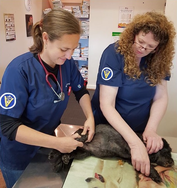
Whenever a dog is lame for more than a day, it is worth the while having the vet check them out. The vet will do a clinical exam and feel up and down (palpate) and manipulate the affected leg to find the origin of the pain. A simple test called the cranial drawer test will be done to evaluate whether the cranial cruciate ligament is intact. While the dog is lying relaxed on his side, the vet will test the stability of the knee by attempting to move the tibia forward whilst stabilising the lower end of the femur. The function of the CCL is to prevent this forward movement of the tibia when weight is placed on the leg, thereby keeping the joint stable. If this ligament is torn, this stabilising mechanism is not present and the tibia will move forward a lot more than what it should. It is important to note that dogs that are tense and painful will guard the joint with tense muscles and this movement is extremely difficult to elicit then. Often you will only be able to feel the cranial drawer sign by doing it under deep sedation. Also, in partial tear cases, the anterior drawer sign may not be pronounced and more difficult to interpret. The veterinarian may request to have these patients admitted for sedation to palpate and examine the leg and also do X-rays while under sedation if required. X-rays are a vital tool in cases where the condition had not been attended to shortly after it happened to evaluate the joint for signs of arthritis secondary to the CCL rupture. With the help of X-rays the vet can also rule out other possible causes of lameness in the hind limbs such as fractures or even bone cancer.
After a diagnosis of a CCL rupture is made, you will have to decide with the help of your veterinarian what course of treatment you are going to follow with your pet. Because of the instability in the joint after a CCL rupture, arthritis will start forming in the joint and will get progressively worse with time. The aim with treatment is to limit arthritis by stabilising the joint and to limit pain as much as possible. Treatment options can be divided into non-surgical (conservative) for cases which were missed and where it may to too late for surgery or surgical for acute cases. To make the decision, the history of the injury, the age and size of the patient as well as the severity of arthritis and degenerative changes in the knee need to be taken into consideration. .
Partial tears could potentially be managed without surgery but most cases, even the ones with partial tears, will require surgery to stabilise the joint. There are many surgical techniques available today and the best will be to discuss with your vet, which method will be the best for your dog. The two main types are intracapsular techniques, where the whole knee joint is opened during the operation, and extra capsular techniques, where the joint capsule is left intact and the operations is done outside of it. Larger dogs usually requires more specialised techniques to give stability. Unfortunately none of the techniques are guaranteed to give a 100 % stability and to prevent arthritis in the long run but it will in most cases prevent the terrible side effects of not doing the surgery at all, leading to excessive connective tissue being formed by the body in an effort to stabilise the knee joint. Aftercare is extremely important as the joint takes time to recover after surgery and owner compliance is of utmost importance. Supportive therapy includes anti-inflammatory drugs for pain management, hydrotherapy, physiotherapy and joint supplementation.
The outcome of surgery is usually very positive with animals making a full recovery in the weeks and months following the operation. Beware though, in large dogs, there is often a scenario where the knee on the opposite side may be affected if the correct aftercare is not taken. Make sure you understand and adhere to the vet’s recommendations to ensure the best possible outcome for your dog.
© 2018 Vetwebsites – The Code Company Trading (Pty.) Ltd.


